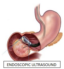
Endoscopic Ultrasound (EUS)
A flexible endoscope which has a small ultrasound device built into the end can be used to see the lining and wall of the esophagus, stomach, small bowel, or colon. The ultrasound component produces sound waves that create visual images of the digestive tract which extend beyond the inner surface lining and also allows visualization of adjacent organs. Endoscopic ultrasound examinations (also called endoluminal endosonography) may be performed through the mouth or through the anus. EUS is performed under sedation.
EUS provides more detailed pictures of the digestive tract anatomy. It can be used to evaluate an abnormality below the surface of the inner lining (mucosa) such as a growth that was detected at a prior endoscopy or by X-ray. EUS, because of its ability to examine the wall layers of the GI tract, provides a detailed picture of the growth, which can help the doctor determine its nature and decide on the best treatment.
Fine Needle Aspiration (FNA) can be performed by passing a biopsy needle down the channel of the endoscope and across the intestinal wall under ultrasound guidance to obtain tissue for the diagnosis and staging of cancer. More recently, EUS has emerged as a therapeutic tool for treating both solid and cystic tumors of the pancreas, alleviating intractable abdominal pain secondary to advanced pancreatic cancer, and obtaining access to the bile ducts and pancreatic duct in cases of failed ERCP.